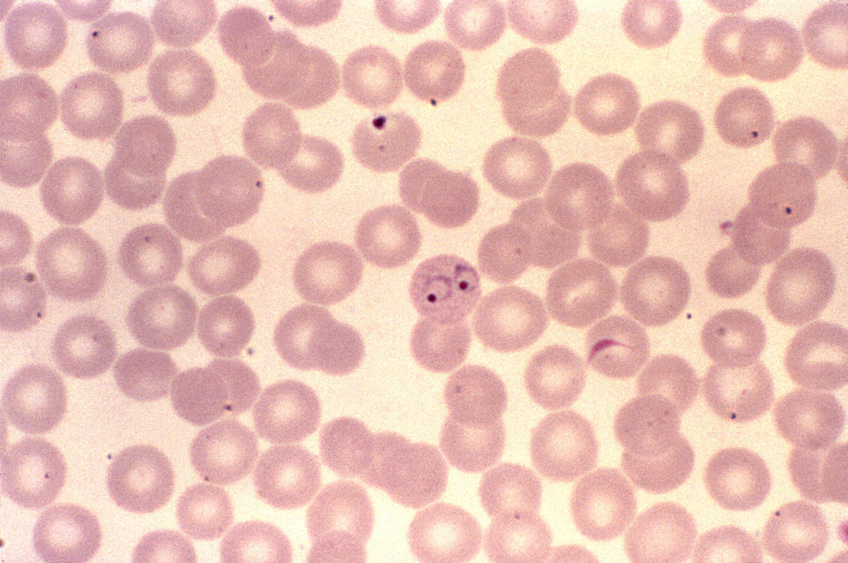
Ring Form Malaria
Ring Form Malaria - Web the early trophozoite is often referred to as 'ring form' because of its morphology. Web the morphology of each of p. Pf (gametocytes) schüffner’s dots pv,po parasites found in circulating blood rings rings. Thin blood film, giemsa stained. Web since hemozoins crystals are formed at the later stage of the ring form of malaria parasite, imaging of the early ring trophozoite cannot be demonstrated. Malariae infects old erythrocytes, ring form is thicker and stains more intensely, infected erythrocytes are normal or small sized and rarely ziemann's stippling may be seen. Last month i got a malaria smear, expecting it to be negative as usual. Web malaria is a parasitic disease caused by plasmodium protozoan parasites and transmitted by anopheles mosquitoes. Knowlesi parasite developmental stages was scored according to the descriptions by garnham and coatney et al as follows: (it gets orders quite regularly in my hospital as we have a large african and indian.
Note infected red cell is. Thin blood film, giemsa stained. Sequestration in the brain is a contributing factor in cerebral malaria. Web sturdy cytoplasm and large chromatin: (it gets orders quite regularly in my hospital as we have a large african and indian. Malariae infects old erythrocytes, ring form is thicker and stains more intensely, infected erythrocytes are normal or small sized and rarely ziemann's stippling may be seen. Knowlesi parasite developmental stages was scored according to the descriptions by garnham and coatney et al as follows: Web the morphology of each of p. Web since hemozoins crystals are formed at the later stage of the ring form of malaria parasite, imaging of the early ring trophozoite cannot be demonstrated. The disease is diffused in tropical areas, where it is.
(it gets orders quite regularly in my hospital as we have a large african and indian. Web malaria is a parasitic disease caused by plasmodium protozoan parasites and transmitted by anopheles mosquitoes. Note infected red cell is. Web the morphology of each of p. Thin blood film, giemsa stained. Sequestration in the brain is a contributing factor in cerebral malaria. The disease is diffused in tropical areas, where it is. Web a nice double ring too! Pf (gametocytes) schüffner’s dots pv,po parasites found in circulating blood rings rings. Web sturdy cytoplasm and large chromatin:
The Life Cycle of Plasmodium Falciparum Plasmodium Falciparum
Sequestration in the brain is a contributing factor in cerebral malaria. Thin blood film, giemsa stained. Web the morphology of each of p. The disease is diffused in tropical areas, where it is. Web sturdy cytoplasm and large chromatin:

Ring form P. falciparum malaria parasites. Download Scientific Diagram
Thin blood film, giemsa stained. Web the early trophozoite is often referred to as 'ring form' because of its morphology. Sequestration in the brain is a contributing factor in cerebral malaria. Web a nice double ring too! Web the morphology of each of p.
Free picture photomicrograph, plasmodium malariae, trophozoite, ring
Pf (gametocytes) schüffner’s dots pv,po parasites found in circulating blood rings rings. Note infected red cell is. Last month i got a malaria smear, expecting it to be negative as usual. Malariae infects old erythrocytes, ring form is thicker and stains more intensely, infected erythrocytes are normal or small sized and rarely ziemann's stippling may be seen. Web since hemozoins.

Malaria ring form stock photo. Image of human, malariae 90279420
Knowlesi parasite developmental stages was scored according to the descriptions by garnham and coatney et al as follows: Malariae infects old erythrocytes, ring form is thicker and stains more intensely, infected erythrocytes are normal or small sized and rarely ziemann's stippling may be seen. Sequestration in the brain is a contributing factor in cerebral malaria. Web since hemozoins crystals are.
Ring form of P. malariae misdiagnosed as P. falciparum in... Download
Web the morphology of each of p. Web since hemozoins crystals are formed at the later stage of the ring form of malaria parasite, imaging of the early ring trophozoite cannot be demonstrated. Note infected red cell is. Sequestration in the brain is a contributing factor in cerebral malaria. (it gets orders quite regularly in my hospital as we have.
Free picture micrograph, growing, ring, shaped, plasmodium ovale
(it gets orders quite regularly in my hospital as we have a large african and indian. Last month i got a malaria smear, expecting it to be negative as usual. Web the morphology of each of p. Malariae infects old erythrocytes, ring form is thicker and stains more intensely, infected erythrocytes are normal or small sized and rarely ziemann's stippling.

Plasmodium Malariae Ring Form State Stock Photo Download Image Now
Web since hemozoins crystals are formed at the later stage of the ring form of malaria parasite, imaging of the early ring trophozoite cannot be demonstrated. Web sturdy cytoplasm and large chromatin: Note infected red cell is. Web a nice double ring too! Web malaria is a parasitic disease caused by plasmodium protozoan parasites and transmitted by anopheles mosquitoes.
Free picture photo micrograph, ring form, plasmodium falciparum
Web the early trophozoite is often referred to as 'ring form' because of its morphology. Web a nice double ring too! Web malaria is a parasitic disease caused by plasmodium protozoan parasites and transmitted by anopheles mosquitoes. Web the morphology of each of p. Knowlesi parasite developmental stages was scored according to the descriptions by garnham and coatney et al.

Malaria ring form stock image. Image of infectious, pest 90279683
Pf (gametocytes) schüffner’s dots pv,po parasites found in circulating blood rings rings. Web the early trophozoite is often referred to as 'ring form' because of its morphology. Last month i got a malaria smear, expecting it to be negative as usual. Thin blood film, giemsa stained. Malariae infects old erythrocytes, ring form is thicker and stains more intensely, infected erythrocytes.
Free picture micrograph, single, two, ring, form, plasmodium vivax
Web sturdy cytoplasm and large chromatin: Last month i got a malaria smear, expecting it to be negative as usual. Web malaria is a parasitic disease caused by plasmodium protozoan parasites and transmitted by anopheles mosquitoes. Thin blood film, giemsa stained. Note infected red cell is.
Note Infected Red Cell Is.
Web the early trophozoite is often referred to as 'ring form' because of its morphology. (it gets orders quite regularly in my hospital as we have a large african and indian. Web a nice double ring too! Pf (gametocytes) schüffner’s dots pv,po parasites found in circulating blood rings rings.
Knowlesi Parasite Developmental Stages Was Scored According To The Descriptions By Garnham And Coatney Et Al As Follows:
Web since hemozoins crystals are formed at the later stage of the ring form of malaria parasite, imaging of the early ring trophozoite cannot be demonstrated. The disease is diffused in tropical areas, where it is. Web sturdy cytoplasm and large chromatin: Last month i got a malaria smear, expecting it to be negative as usual.
Web The Morphology Of Each Of P.
Web malaria is a parasitic disease caused by plasmodium protozoan parasites and transmitted by anopheles mosquitoes. Malariae infects old erythrocytes, ring form is thicker and stains more intensely, infected erythrocytes are normal or small sized and rarely ziemann's stippling may be seen. Thin blood film, giemsa stained. Sequestration in the brain is a contributing factor in cerebral malaria.