How To Read A Ultrasound Report
How To Read A Ultrasound Report - Web it is at this time that the sonographer will measure the size of your baby, check the major organs, measure the level of amniotic fluid to make sure that it's right, and check the position of the placenta. Web knowing how to read a pregnancy ultrasound. Pet or pet/ct second opinion; Web abdominal ultrasound can diagnose many causes of abdominal pain. Besides, learn how to know it is a girl or boy by reading an ultrasound. During the 12th week, the ultrasound. The test helps your health care team find out: Web learning how to read an ultrasound will help you get important details about your baby from the picture. There are many reasons you might have an ultrasound. Web how to read your abdominal ultrasound report your healthcare provider (usually a doctor, nurse practitioner, or physician assistant) sometimes uses medical imaging tests to diagnose and treat diseases.
Web learning how to read an ultrasound will help you get important details about your baby from the picture. Web abdominal ultrasound can diagnose many causes of abdominal pain. Transvaginal ultrasound ‘transvaginal’ means through the vagina. The test helps your health care team find out: Web this article summarises the best practice in reporting of ultrasound examinations based on international literature and addresses key topics including report structure, clinical content, style and. The size and shape of your. Web an echo test can allow your health care team to look at your heart’s structure and check how well your heart functions. It is generally done when the fetus is below 10 weeks. Web background and objectives the transversus abdominis plane block (tap) can be applied using different approaches, resulting in varying cutaneous analgesic distributions. Researchers have mapped out the expected measurements for specific points in early pregnancy, an early ultrasound.
Besides, learn how to know it is a girl or boy by reading an ultrasound. Web learning how to read an ultrasound will help you get important details about your baby from the picture. After watching this video you will be able to understand the basic terms that are. Web background and objectives the transversus abdominis plane block (tap) can be applied using different approaches, resulting in varying cutaneous analgesic distributions. This study aimed to assess the cutaneous sensory block area (csba) after ultrasound. Web it is at this time that the sonographer will measure the size of your baby, check the major organs, measure the level of amniotic fluid to make sure that it's right, and check the position of the placenta. Web the radiology report represents the sum of a radiologist’s highest level of synthesis and insight into a patient’s condition. Web this article summarises the best practice in reporting of ultrasound examinations based on international literature and addresses key topics including report structure, clinical content, style and. During the 12th week, the ultrasound. A radiologist is a doctor who supervises these exams, reads and interprets the images, and writes a report.

Confirmed Scans Gallery The Gender Experts Ultrasound gender
The size and shape of your. Web background and objectives the transversus abdominis plane block (tap) can be applied using different approaches, resulting in varying cutaneous analgesic distributions. Pet or pet/ct second opinion; Web knowing how to read a pregnancy ultrasound. Besides, learn how to know it is a girl or boy by reading an ultrasound.
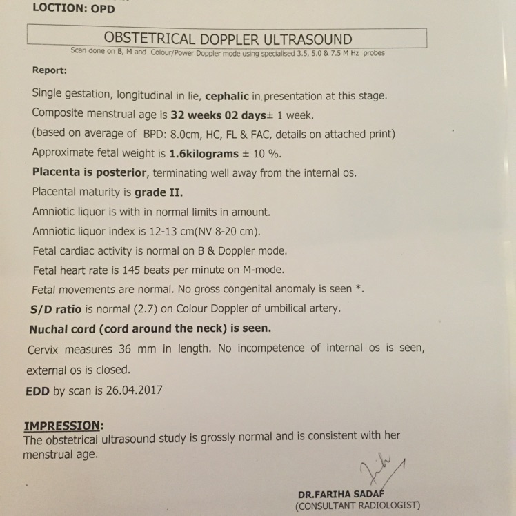
Is this ultrasound report is normal? Glow Community
Web background and objectives the transversus abdominis plane block (tap) can be applied using different approaches, resulting in varying cutaneous analgesic distributions. The test helps your health care team find out: This study aimed to assess the cutaneous sensory block area (csba) after ultrasound. Web abdominal ultrasound can diagnose many causes of abdominal pain. Web the radiology report represents the.

how to read an ultrasound report // normal report // in hindi // part
Web knowing how to read a pregnancy ultrasound. There are many reasons you might have an ultrasound. If you are going for the ultrasound test during the 8th week of pregnancy, then your fetus will be similar to the size of a single baked bean. Besides, learn how to know it is a girl or boy by reading an ultrasound..

Whole Abdomen Ultrasound In Hindi fitriblog1
Researchers have mapped out the expected measurements for specific points in early pregnancy, an early ultrasound. If you are going for the ultrasound test during the 8th week of pregnancy, then your fetus will be similar to the size of a single baked bean. 👉do subscribe now👈👇for any doubt & query👇👉follow me on instagram : The size and shape of.
How Do You Read An Ultrasound Image Images Poster
Pet or pet/ct second opinion; Web nursing/neet/aiims/jipmer lectures, books, question papers, notes, pdf. There are many reasons you might have an ultrasound. Web abdominal ultrasound can diagnose many causes of abdominal pain. Web an echo test can allow your health care team to look at your heart’s structure and check how well your heart functions.

How To Find Out Gender Of Baby In Ultrasound Report Baby Viewer
Web hello friends in this video, i have explained how you can read your own pregnancy ultrasound report easily. Transvaginal ultrasound ‘transvaginal’ means through the vagina. There are many reasons you might have an ultrasound. This study aimed to assess the cutaneous sensory block area (csba) after ultrasound. Web nursing/neet/aiims/jipmer lectures, books, question papers, notes, pdf.

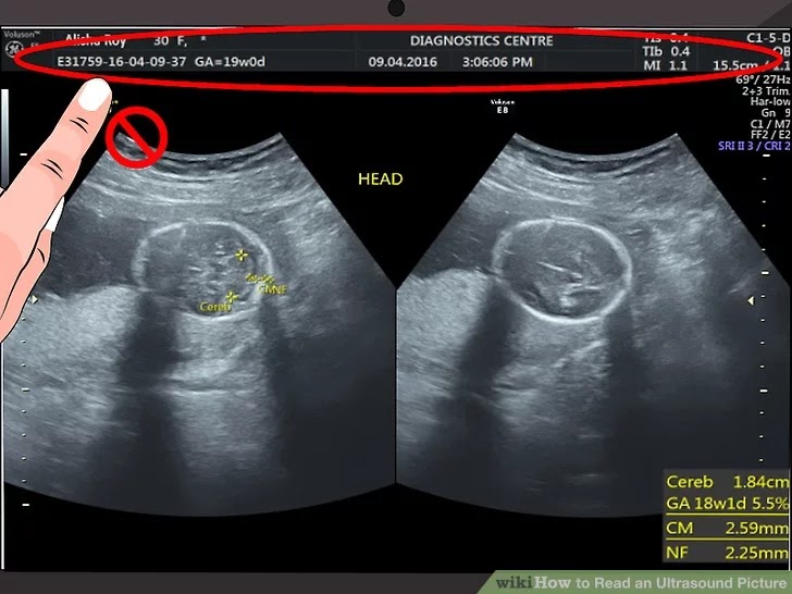
How to Read an Ultrasound Picture 8 Steps (with Pictures)
Transvaginal ultrasound ‘transvaginal’ means through the vagina. 👉do subscribe now👈👇for any doubt & query👇👉follow me on instagram : Web learning how to read an ultrasound will help you get important details about your baby from the picture. Web abdominal ultrasound can diagnose many causes of abdominal pain. Web knowing how to read a pregnancy ultrasound.
28+ How To Read Ultrasound Picture KnishaAshtin
Transvaginal ultrasound ‘transvaginal’ means through the vagina. Pet or pet/ct second opinion; Web knowing how to read a pregnancy ultrasound. If you are going for the ultrasound test during the 8th week of pregnancy, then your fetus will be similar to the size of a single baked bean. It is generally done when the fetus is below 10 weeks.

Level 2 ultrasound रिपोर्ट देखना सीखे How To Read Ultrasound Report
Web knowing how to read a pregnancy ultrasound. Web hello friends in this video, i have explained how you can read your own pregnancy ultrasound report easily. Web background and objectives the transversus abdominis plane block (tap) can be applied using different approaches, resulting in varying cutaneous analgesic distributions. There are many reasons you might have an ultrasound. Web abdominal.

Level 2 ultrasound रिपोर्ट देखना सीखे How To Read Ultrasound Report
Web knowing how to read a pregnancy ultrasound. After watching this video you will be able to understand the basic terms that are. If you are going for the ultrasound test during the 8th week of pregnancy, then your fetus will be similar to the size of a single baked bean. Web learning how to read an ultrasound will help.
It Is The Most Important Product That Radiologists Generate To Help Direct.
Web learning how to read an ultrasound will help you get important details about your baby from the picture. Web expected results in early pregnancy. Web knowing how to read a pregnancy ultrasound. 👉do subscribe now👈👇for any doubt & query👇👉follow me on instagram :
Pet Or Pet/Ct Second Opinion;
It is generally done when the fetus is below 10 weeks. Web this article summarises the best practice in reporting of ultrasound examinations based on international literature and addresses key topics including report structure, clinical content, style and. The size and shape of your. Providers use kidney ultrasound to assess the size, location and shape of your kidneys and related structures, such as your ureters and bladder.
Web An Echo Test Can Allow Your Health Care Team To Look At Your Heart’s Structure And Check How Well Your Heart Functions.
During the 12th week, the ultrasound. This study aimed to assess the cutaneous sensory block area (csba) after ultrasound. Transvaginal ultrasound ‘transvaginal’ means through the vagina. The test helps your health care team find out:
Web Background And Objectives The Transversus Abdominis Plane Block (Tap) Can Be Applied Using Different Approaches, Resulting In Varying Cutaneous Analgesic Distributions.
If you are going for the ultrasound test during the 8th week of pregnancy, then your fetus will be similar to the size of a single baked bean. A radiologist is a doctor who supervises these exams, reads and interprets the images, and writes a report. Researchers have mapped out the expected measurements for specific points in early pregnancy, an early ultrasound. Web nursing/neet/aiims/jipmer lectures, books, question papers, notes, pdf.